Table of Contents
Lung cancer can now be diagnosed and treated at the same time using a new type of robotic surgery. Margaret Kirkham, 77, a retired career civil servant from Chiswick, west London, was the first patient in the world to undergo the new two-in-one procedure, she tells ADRIAN MONTI.
The patient
A little over two years ago, I suddenly became out of breath and even had to stop talking mid-sentence to catch my breath. My GP examined me and sent me to the emergency room for tests, including a chest x-ray.
The next morning they called me to come back immediately for a CT scan as it looked like part of my lung had collapsed. I was quite surprised.
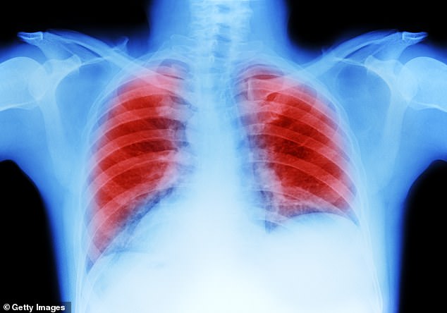
A doctor explained to me that they had found a large mass in my left lung, which looked like cancer. Luckily, they told me, it had not spread.
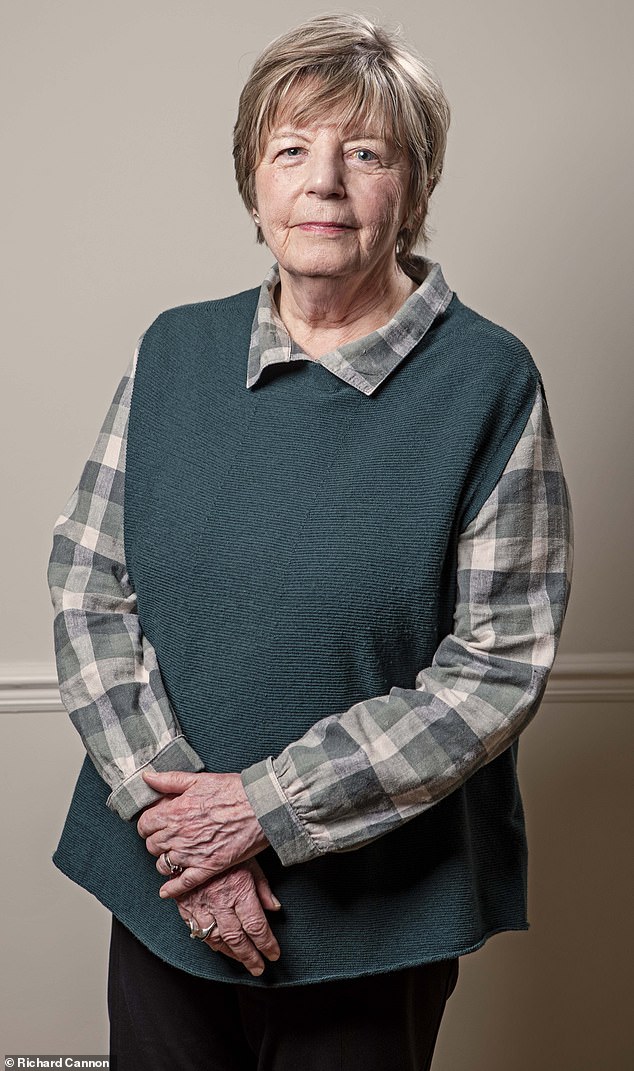
Margaret Kirkham, 77, was the first patient in the world to be treated with the 2-in-1 method
I used to be a smoker, but I had quit 20 years earlier, and although I was told that my cancer was probably not caused by tobacco due to its molecular structure, I still felt guilty.
My tumor measured 7 cm by 6 cm by 6 cm: I imagined it as a large solid rectangle. It couldn’t be removed surgically because it was close to my heart and entwined in the main airways to the lung. This sounded scary, but my doctor assured me that it was growing slowly and could be controlled with treatment.
In April 2022 I started chemotherapy and radiotherapy and it worked quickly, my lungs re-inflated which made breathing much easier.
A scan in July showed the tumor had shrunk dramatically. Then I received monthly immunotherapy until last August. By then, the original tumor had disappeared, but there was a small new nodule in my left lung. However, it was not safe to have more radiotherapy and I could not have surgery.
But my consultant mentioned that a new technique was being tested for smaller, harder-to-reach lung tumors. This was ablation, in which heat is used to destroy the cancer. I was referred to Professor Pallav Shah, the lung specialist responsible for the trial.
He explained that they would give me general anesthesia and then they would pass a flexible tube down my windpipe. Connected to a sophisticated robotic system, it could navigate deep into my lungs to take a biopsy and then they would use a small ablation tool to use heat to destroy small cancerous nodules.
After surgery in November, I felt good and pain-free.
It made me smile knowing that I was the first to undergo this procedure. I’ve never been first at anything before!
Scans showed the tumor was successfully removed and I was back to my normal life within a week. I have slight shortness of breath due to scar tissue from the ablation and radiation, but it’s a small price to pay.
I am not taking any medication and I feel good and positive and am making up for lost time. I was just on a painting trip to Cádiz. I am very grateful to have participated in this trial, which I hope will benefit many others.
The specialist
Professor Pallav Shah is a consultant respiratory health physician at the Royal Brompton Hospital, London.
Lung cancer is the third most common cancer in the UK, with 40,000 cases diagnosed annually and a very low survival rate. (Only 20 percent of patients live five years or more.)
This is because it does not cause obvious symptoms, so it is usually detected at an advanced stage, often during investigations for symptoms such as a persistent cough, weight loss or chest pain. But if we can treat tumors that are less than 10 mm, the cure rate is 92 percent.
Currently, most people are diagnosed when the cancer spot or nodule measures more than 30 mm, where the cure rate is 68 percent.
Since April last year, we have been using a pioneering robot-assisted system, the Ion Endoluminal System, at the Royal Brompton. Using a robotically controlled catheter system, it can reach nodules up to 6 mm.
This allows us to perform a minimally invasive biopsy in any part of the lung, even in places that are normally very difficult to access within the tiny bronchioles or branches of the airway “tree.”

Lung cancer is the third most common cancer in the UK, with 40,000 cases diagnosed annually
We can now also use the Ion system with a new ablation tool, which uses heat to destroy cancer, in patients who are not suitable for conventional surgery or radiotherapy.
Currently, a standard lung cancer biopsy involves inserting a needle through the chest wall into the lung, using a CT scanner as a guide.
This is done under general anesthesia and results take a week. There is a risk of lung puncture (this occurs in 25 percent of cases), which can lead to bleeding, clots, strokes and even death. It is not appropriate for patients with poor lung function (for example, those with severe emphysema) or nodules in a difficult location.
The robotic approach, also performed under general anesthesia, has a less than 10 percent risk of pneumothorax—lung collapse. We do a biopsy to verify that the nodule is cancerous before performing the ablation.
First, a CT scan of the lungs is uploaded to the Ion system, providing a highly detailed 3D roadmap of the inside of the lungs. The system automatically traces a route to the node.
An ultra-thin, flexible tube (catheter) is then passed into the patient’s airway through the mouth, while we monitor the progress on a screen. Once the catheter reaches the nodule, the needle is deployed to collect a tissue sample.
Not only does it allow us to target smaller nodules with greater precision, but the robotic catheter is much more flexible than the conventional bronchoscope (a flexible camera used to examine the airways).
The bronchoscope can only reach 65 percent of points smaller than 20 mm, but the robotic system reaches more than 90 percent of spots smaller than 10 mm.
Now we can also treat the patient during the same session.
Margaret was the first patient in the world to have a biopsy and robotic microwave ablation performed in a single 45-minute procedure, saving her 30 to 45 minutes of subsequent ablation treatment.
This is due to another technological advance: a new type of ablation tool called MicroBlate Flex, developed by the British company Creo, which is only 1.8 mm in diameter.
This goes through the same robotic catheter that the biopsy was done through, so the exact same site is biopsied and treated. This cannot be done with standard ablation tools, which must pass through the chest wall.
Margaret’s lung nodule was less than 10mm, but we also performed a margin ablation to ensure all cancerous tissue was destroyed. The ablation took three minutes.
The beauty of microwave energy is that we can repeat it if necessary, which is not the case with radiotherapy, where only a certain amount can be delivered due to tissue damage.
Performing two procedures at once is a game-changer, saving time for both doctors and patients, who also save a delay between biopsy and ablation, during which their cancer continues to grow.
Patients in the trial spend the night in hospital, but in the future it should be a daytime procedure. Our goal is to use it to treat small, early cancers in patients who are not suitable for surgery; For example, they may also have heart disease.
Although we need to send biopsy samples taken with the Ion for analysis, which can take a week, in the future we hope to use AI to analyze them in a few minutes, allowing us to perform ablation immediately.
In Margaret’s case, we were already sure that her nodule was cancerous (due to previous treatment), so we did a biopsy followed immediately by ablation to demonstrate that the two-in-one technique could be performed successfully.
So far we have performed ablation in nine patients, without serious side effects.

