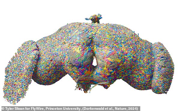
The structure of the brain is one of the most perplexing and complex enigmas in the universe.
But now, an international team of scientists has created the first map showing every neuron and connection in an adult brain.
This ‘wiring diagram’, created by the FlyWire Consortium, reveals each of the 139,255 neurons in a fruit fly brain and the 50 million connections between them.
While the human brain has about a million times more neurons than a fly’s, researchers say this brings us closer to understanding our own mind.
Project co-leader Dr Gregory Jefferis from the University of Cambridge said: “Brain wiring diagrams are a first step in understanding everything we care about: how we control our movement, answer the phone or recognize a friend. “. ‘
An international team of scientists has created the first map showing every neuron and connection in an adult brain.
Although the brain of an adult fruit fly measures less than a millimeter in diameter, it is still an enormously complicated structure to study.
To produce this innovative map, the brain of an adult fruit fly was carefully cut into 7,000 segments, each just 40 nanometers thick.
Each segment was then scanned individually using a high-powered electron microscope to reveal the individual cells that make up the brain.
The resulting data set occupied 100 Terabytes of storage, the equivalent of 2,500 high-definition movies.
The researchers developed an AI capable of reconstructing a map of the brain, identifying each and every one of the neurons and connections.
However, since the AI was still prone to making some errors, a team of 287 researchers from more than 76 laboratories around the world reviewed the entire data set to check for errors.
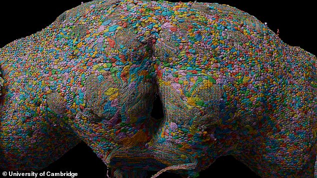
The map contains the location of 139,255 neurons and 50 million connections between them.
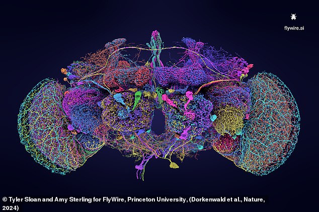
The researchers cut a fly’s brain into 7,000 slices and scanned them with an electron microscope to reveal how each neuron is connected throughout the brain.
If one person had worked non-stop to check the data, researchers estimate it would have taken 33 years to complete the project.
While the effort was monumental, the reward is the most detailed map of any animal’s brain ever produced.
This map has been published in two articles in Nature and has been made available to other scientists.
Compared to previous attempts to detail small regions of a fly’s brain, this new map contains seven times as many neurons and records 54.5 million individual connections.
Previously, the largest brains that had been completely mapped belonged to fruit fly larvae, which have 3,016 neurons, or nematodes, which only have 302.
This is the first time scientists have been able to map the brain of an animal that can walk, see and engage in complex behaviors.
The researchers believe this could pave the way to understanding the fundamental dynamics that enable complex behavior.
This map reveals the connections within the brain in never-before-seen detail and contains seven times more neurons than previous maps. This image shows different cells color coded according to the chemicals they use to transfer information.
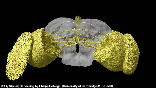
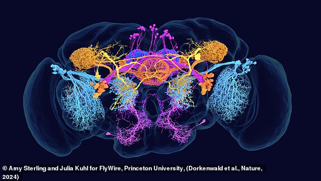
The map also allows researchers to study the brain regions responsible for different skills. For example, this image shows the visual system of a fly.
Dr Jefferis says: “Flies can do all sorts of complicated things like walk, fly, navigate and the males sing to the females.
“If we want to understand how the brain works, we need a mechanistic understanding of how all the neurons fit together and allow us to think.”
One idea that has already emerged from the study is that our brains may not be as unique as we think.
Compared to previous partial brain maps, the researchers discovered significant similarities in how neurons were connected.
This suggests that our brain may not be “a single structure like a snowflake,” but rather follows set patterns.
The researchers found that only 0.5 percent of the brain’s neurons had variations that caused them to be wired differently.
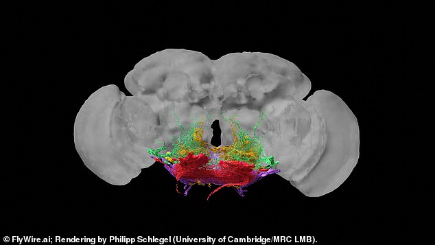
This map shows the 100 neurons that make up a fly’s motor system. This is the first time researchers have mapped the brain of an animal capable of walking and seeing.
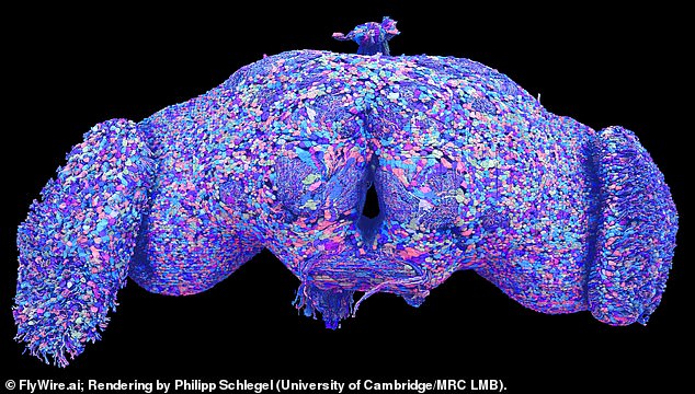
Although a fly brain contains a million times fewer neurons than the human brain, researchers hope this could lay the groundwork for studying the brains of larger organisms.
These poorly connected neurons could be the source of our mind’s individuality or brain disorders.
However, seeing how neurons fit together is only the first part of the puzzle.
If scientists want to start digitally simulating the fruit fly brain, we also need to know what all parts of the brain are doing.
Co-lead author Dr Philipp Schlegel, from the MRC Laboratory of Molecular Biology, says: “This data set is a bit like Google Maps, but for brains: the raw wiring diagram between neurons is like knowing which structures in Satellite images of the Earth correspond to streets and buildings.
“Annotating neurons is like adding street and city names, business opening hours, phone numbers, reviews, etc. to a map; you need both to make it really useful.”
Researchers have already identified more than 8,400 unique cell types responsible for abilities such as sight or movement, including 4,581 previously unknown types.
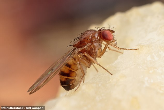
Since fruit flies are capable of complex behaviors, by mapping their brains researchers can learn more about the neural circuits that make this possible.
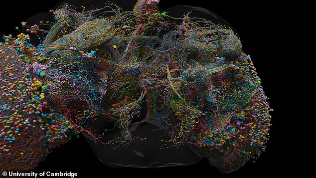
The next goal will be to identify the role of individual neurons within the map (pictured) so researchers can begin simulating brains digitally.
These allow us to see, neuron by neuron in detail, the structures responsible for a fly’s natural navigation skills, their ability to recognize shapes, and even how they listen to other flies’ songs.
Since fruit flies are a common animal in research laboratories around the world, researchers believe this knowledge will lead to a better understanding of the inner workings of the brain.
Co-lead researcher Professor David Bock, from the University of Vermont, says: ‘This will inevitably lead to a deeper understanding of how nervous systems process, store and remember information.
“I believe this approach points the way forward for the analysis of future connectomes of the entire brain, both in the fly and in other species.”


